- 2-in-1 system - High end radiography and fluoroscopy
-
2-in-1 system - High end radiography and fluoroscopy
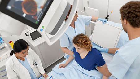
Philips CombiDiagnost R90 premium cross-functional system is a true all around performer. Applications include chest, spine, upper and lower extremities, skull, as well as gastro-intestinal exams, arthrography, venography, lymphography, myelography and Digital Subtraction Angiography (DSA). - Flexible geometry - Versatility in use
-
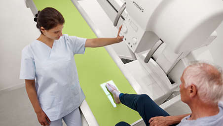
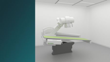
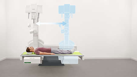
Flexible geometry - Versatility in use
The remote controlled tiltable table (-90° to +90°) is ideal for all standard fluoroscopy studies and for traditional X-rays including image stitching. A tilting tube column mechanism enables angled projections in any table position. The tabletop can hold a patient weighing up to 284 kg (626 lbs.) without restricting movement. The intuitive touchscreen controls geometry movements and fluoroscopy parameters. - Eleva Tube Head¹ - Patient centered work
-
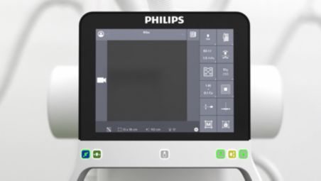
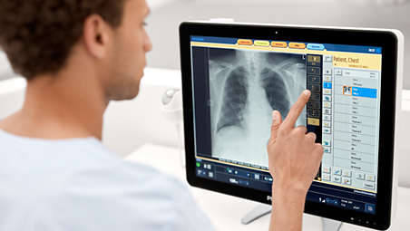
Eleva Tube Head¹ - Patient centered work
When equipped with a ceiling suspension CombiDiagnost R90 features the intuitive Eleva Tube Head, a modern smart touch interface in the examimation room. It allows the user to quickly change the most vital parameters for rad imaging directly at the tube head. The Eleva Tube Head offers an optional live camera image for improved positioning support. - Grid-Controlled Fluoroscopy - Fully automatic dose adjustment
-
Grid-Controlled Fluoroscopy (GCF) - Fully automatic dose adjustment
GCF is an exclusive Philips technology of pulsed fluoroscopy, providing superb image quality at low dose. Dose management is achieved by the use of a grid-switch mechanism in the X-ray tube. Moreover, X-ray parameters kV, mA and time are controlled within each single pulse (in-pulse control). - Dynamic UNIQUE image processing - Hidden details revealed
-
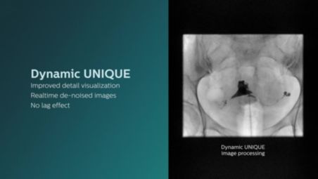
Dynamic UNIQUE image processing - Hidden details revealed
Dynamic UNIQUE image processing software delivers consistently uniform clinical image quality for all anatomic regions within one image by automatically adjusting the balance between overexposed and underexposed areas. - UNIQUE 2¹ - Next generation of rad image processing
-
UNIQUE 2¹ - Next generation of rad image processing
UNIQUE 2 image processing delivers fast, outstanding digital rad images. It significantly improves image quality like homogeneous black backgrounds, reduced noise, and automatic enhancement of small details. - Stitching - Full leg and full spine imaging
-
Stitching - Full leg and full spine imaging
The optional automatic image stitching software allows to acquire long-length images. Image acquisition is possible on the table in every position from horizontal to upright¹ and at the vertical stand. The algorithm works fully automatically. A set of smart accessories provides excellent patient comfort and superb image quality. - Digital Subtraction Angiography¹ - High quality vessel imaging
-
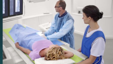
Digital Subtraction Angiography¹ - High quality vessel imaging
With Philips Digital Subtraction Angiography (DSA), blood vessels can be visiualized at UNIQUE image quality. Automatic examination pre-sets support a smooth and efficient workflow in angiography examinations. - SkyPlates - Detectors that deliver
-
SkyPlates - Detectors that deliver
CombiDiagnost R90’s large and small SkyPlate wireless portable digital detectors are lightweight to allow comfortable positioning at the system. They pair nicely with our SkyFlow Plus intelligent software for non-grid bedside exams and can be shared between compatible systems. - SkyFlow Plus - Grid-like contrast for free rad examinations
-
SkyFlow Plus - Grid-like contrast for free rad examinations
SkyFlow Plus is the industry’s first scatter radiation correction algorithm for portable X-rays. When doing DR exams without a grid, Philips SkyFlow Plus produces images with grid-like contrast. The software reduces the effect of scattered radiation for non-grid free exposures. - Eleva user interface - Quick, intuitive workflow
-
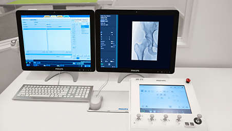
Eleva user interface - Quick, intuitive workflow
The intuitive Eleva user interface provides all the tools and controls necessary for seamless procedures. This common platform is harmonized across the Philips radiography and fluoroscopy portfolio. It is easy to learn and use and therefore reduces training effort. - Philips Bone Suppression¹ - Improved nodule detection
-
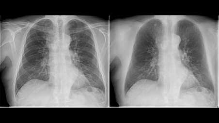
Philips Bone Suppression¹ - Improved nodule detection
Philips Bone Suppression² software helps remove bone structures from adult erect chest images acquired at the vertical stand for an unobstructed view of soft tissue. This clear view can help to ensure a more accurate image interpretation. As part of Philips’ Eleva platform, Bone Suppression is integrated into the regular system workflow.
2-in-1 system - High end radiography and fluoroscopy
2-in-1 system - High end radiography and fluoroscopy
2-in-1 system - High end radiography and fluoroscopy
Flexible geometry - Versatility in use
Flexible geometry - Versatility in use
Flexible geometry - Versatility in use
Eleva Tube Head¹ - Patient centered work
Eleva Tube Head¹ - Patient centered work
Eleva Tube Head¹ - Patient centered work
Grid-Controlled Fluoroscopy (GCF) - Fully automatic dose adjustment
Grid-Controlled Fluoroscopy (GCF) - Fully automatic dose adjustment
Grid-Controlled Fluoroscopy (GCF) - Fully automatic dose adjustment
Dynamic UNIQUE image processing - Hidden details revealed
Dynamic UNIQUE image processing - Hidden details revealed
Dynamic UNIQUE image processing - Hidden details revealed
UNIQUE 2¹ - Next generation of rad image processing
UNIQUE 2¹ - Next generation of rad image processing
UNIQUE 2¹ - Next generation of rad image processing
Stitching - Full leg and full spine imaging
Stitching - Full leg and full spine imaging
Stitching - Full leg and full spine imaging
Digital Subtraction Angiography¹ - High quality vessel imaging
Digital Subtraction Angiography¹ - High quality vessel imaging
Digital Subtraction Angiography¹ - High quality vessel imaging
SkyPlates - Detectors that deliver

SkyPlates - Detectors that deliver
SkyPlates - Detectors that deliver
SkyFlow Plus - Grid-like contrast for free rad examinations
SkyFlow Plus - Grid-like contrast for free rad examinations
SkyFlow Plus - Grid-like contrast for free rad examinations
Eleva user interface - Quick, intuitive workflow
Eleva user interface - Quick, intuitive workflow
Eleva user interface - Quick, intuitive workflow
Philips Bone Suppression¹ - Improved nodule detection
Philips Bone Suppression¹ - Improved nodule detection
Philips Bone Suppression¹ - Improved nodule detection
- 2-in-1 system - High end radiography and fluoroscopy
- Flexible geometry - Versatility in use
- Eleva Tube Head¹ - Patient centered work
- Grid-Controlled Fluoroscopy - Fully automatic dose adjustment
- 2-in-1 system - High end radiography and fluoroscopy
-
2-in-1 system - High end radiography and fluoroscopy
Philips CombiDiagnost R90 premium cross-functional system is a true all around performer. Applications include chest, spine, upper and lower extremities, skull, as well as gastro-intestinal exams, arthrography, venography, lymphography, myelography and Digital Subtraction Angiography (DSA). - Flexible geometry - Versatility in use
-
Flexible geometry - Versatility in use
The remote controlled tiltable table (-90° to +90°) is ideal for all standard fluoroscopy studies and for traditional X-rays including image stitching. A tilting tube column mechanism enables angled projections in any table position. The tabletop can hold a patient weighing up to 284 kg (626 lbs.) without restricting movement. The intuitive touchscreen controls geometry movements and fluoroscopy parameters. - Eleva Tube Head¹ - Patient centered work
-
Eleva Tube Head¹ - Patient centered work
When equipped with a ceiling suspension CombiDiagnost R90 features the intuitive Eleva Tube Head, a modern smart touch interface in the examimation room. It allows the user to quickly change the most vital parameters for rad imaging directly at the tube head. The Eleva Tube Head offers an optional live camera image for improved positioning support. - Grid-Controlled Fluoroscopy - Fully automatic dose adjustment
-
Grid-Controlled Fluoroscopy (GCF) - Fully automatic dose adjustment
GCF is an exclusive Philips technology of pulsed fluoroscopy, providing superb image quality at low dose. Dose management is achieved by the use of a grid-switch mechanism in the X-ray tube. Moreover, X-ray parameters kV, mA and time are controlled within each single pulse (in-pulse control). - Dynamic UNIQUE image processing - Hidden details revealed
-
Dynamic UNIQUE image processing - Hidden details revealed
Dynamic UNIQUE image processing software delivers consistently uniform clinical image quality for all anatomic regions within one image by automatically adjusting the balance between overexposed and underexposed areas. - UNIQUE 2¹ - Next generation of rad image processing
-
UNIQUE 2¹ - Next generation of rad image processing
UNIQUE 2 image processing delivers fast, outstanding digital rad images. It significantly improves image quality like homogeneous black backgrounds, reduced noise, and automatic enhancement of small details. - Stitching - Full leg and full spine imaging
-
Stitching - Full leg and full spine imaging
The optional automatic image stitching software allows to acquire long-length images. Image acquisition is possible on the table in every position from horizontal to upright¹ and at the vertical stand. The algorithm works fully automatically. A set of smart accessories provides excellent patient comfort and superb image quality. - Digital Subtraction Angiography¹ - High quality vessel imaging
-
Digital Subtraction Angiography¹ - High quality vessel imaging
With Philips Digital Subtraction Angiography (DSA), blood vessels can be visiualized at UNIQUE image quality. Automatic examination pre-sets support a smooth and efficient workflow in angiography examinations. - SkyPlates - Detectors that deliver
-
SkyPlates - Detectors that deliver
CombiDiagnost R90’s large and small SkyPlate wireless portable digital detectors are lightweight to allow comfortable positioning at the system. They pair nicely with our SkyFlow Plus intelligent software for non-grid bedside exams and can be shared between compatible systems. - SkyFlow Plus - Grid-like contrast for free rad examinations
-
SkyFlow Plus - Grid-like contrast for free rad examinations
SkyFlow Plus is the industry’s first scatter radiation correction algorithm for portable X-rays. When doing DR exams without a grid, Philips SkyFlow Plus produces images with grid-like contrast. The software reduces the effect of scattered radiation for non-grid free exposures. - Eleva user interface - Quick, intuitive workflow
-
Eleva user interface - Quick, intuitive workflow
The intuitive Eleva user interface provides all the tools and controls necessary for seamless procedures. This common platform is harmonized across the Philips radiography and fluoroscopy portfolio. It is easy to learn and use and therefore reduces training effort. - Philips Bone Suppression¹ - Improved nodule detection
-
Philips Bone Suppression¹ - Improved nodule detection
Philips Bone Suppression² software helps remove bone structures from adult erect chest images acquired at the vertical stand for an unobstructed view of soft tissue. This clear view can help to ensure a more accurate image interpretation. As part of Philips’ Eleva platform, Bone Suppression is integrated into the regular system workflow.
2-in-1 system - High end radiography and fluoroscopy
2-in-1 system - High end radiography and fluoroscopy
2-in-1 system - High end radiography and fluoroscopy
Flexible geometry - Versatility in use
Flexible geometry - Versatility in use
Flexible geometry - Versatility in use
Eleva Tube Head¹ - Patient centered work
Eleva Tube Head¹ - Patient centered work
Eleva Tube Head¹ - Patient centered work
Grid-Controlled Fluoroscopy (GCF) - Fully automatic dose adjustment
Grid-Controlled Fluoroscopy (GCF) - Fully automatic dose adjustment
Grid-Controlled Fluoroscopy (GCF) - Fully automatic dose adjustment
Dynamic UNIQUE image processing - Hidden details revealed
Dynamic UNIQUE image processing - Hidden details revealed
Dynamic UNIQUE image processing - Hidden details revealed
UNIQUE 2¹ - Next generation of rad image processing
UNIQUE 2¹ - Next generation of rad image processing
UNIQUE 2¹ - Next generation of rad image processing
Stitching - Full leg and full spine imaging
Stitching - Full leg and full spine imaging
Stitching - Full leg and full spine imaging
Digital Subtraction Angiography¹ - High quality vessel imaging
Digital Subtraction Angiography¹ - High quality vessel imaging
Digital Subtraction Angiography¹ - High quality vessel imaging
SkyPlates - Detectors that deliver
SkyPlates - Detectors that deliver
SkyPlates - Detectors that deliver
SkyFlow Plus - Grid-like contrast for free rad examinations
SkyFlow Plus - Grid-like contrast for free rad examinations
SkyFlow Plus - Grid-like contrast for free rad examinations
Eleva user interface - Quick, intuitive workflow
Eleva user interface - Quick, intuitive workflow
Eleva user interface - Quick, intuitive workflow
Philips Bone Suppression¹ - Improved nodule detection
Philips Bone Suppression¹ - Improved nodule detection
Philips Bone Suppression¹ - Improved nodule detection
Specifications
- Generator
-
Generator Power - 65 kW, 80kW optional
Tube voltage exposure - 40 - 150 kV
Exposure techniques - Manual or Automatic Exposure Control kV-mA-s - Intelligent Exposure (IQX)
Tube voltage flouroscopy - 40 - 125 kV
Fluoroscopy techniques - Continous, Pulsed fluoroscopy, Grid-controlled fluoroscopy (option)
-
- Applicational Range
-
Applicational Range Fluoroscopy - Gastro intestinal, urinary studies, ERCP, fluoroscopy guided injections etc
Radiography - Skull, ribs, spine, pelvis, upper extremities, long spine, lung, abdomen etc
-
- Geometry
-
Geometry Table tilt angle - -90°/+90°
Motorized compressor - 3 kg - 15 kg (6.6 lbs - 33.1 lbs) compression force
Tube column movement range - 160 cm (63")
Source to Image Distance (SID) - 113 cm – 183 cm (44" to 72")
Maximum patient weight - 284 kg (626 lbs) without limitations
Table height - 65 cm - 133 cm ( 25’’ – 52.2’’)
-
- Tube: SRM 0608 ROT GS 505 (GCF)
-
Tube: SRM 0608 ROT GS 505 (GCF) Anode heat storage capacity - 800 kHU (593kJ)
Focal spot - 0.6 /0.8
Maximum voltage - 125 kV (110 kV in GCF)
-
- Tube: SRO 33100 ROT380 (PCF)
-
Tube: SRO 33100 ROT380 (PCF) Anode heat storage capacity - 300 kHU (200kJ)
Focal spot - 0.6 /1.2
Maximum voltage - 150 kV
-
- Dynamic Flat Detector
-
Dynamic Flat Detector Detector Size - 43 cm x 43 cm (17"x17")
Image matrix size - 2,874 pixel x 2,840 pixel
Pixel size - 148 µm
Acquisition mode PCF - Up to 6 fps
Acquisition mode GCF - Up to 30 fps
-
- Tube: SRO 33100 ROT380 (for CS)
-
Tube: SRO 33100 ROT380 (for CS) Anode heat storage capacity - 300 kHU (200kJ)
Focal spot - 0.6 /1.2
Maximum voltage - 150 kV
-
- Eleva Tube Head
-
Eleva Tube Head Control buttons - color coded control buttons; capacitive sensor for three-axis brake-release
Data displayed includes - Patient data, preview images, generator settings, live camera image (option)
Full color LCD touch display - 30.7 cm (12.1")
-
- Wireless detector: SkyPlate Large
-
Wireless detector: SkyPlate Large Type - Digital Csl (Cesium Iodide) flat detector
Detector size - 35 cm x 43 cm (14" x 17")
Image matrix size - 2330 x 2846 pixel
Active area - 34.48 cm x 42.12 cm (13.6" x 16.6")
-
- Wireless detector: SkyPlate Small
-
Wireless detector: SkyPlate Small Image matrix size - 1500 x 1920 pixel
Active area - 22.2 cm x 28.4 cm (8.7" x 11.2")
Type - Digital Csl (Cesium Iodide) flat detector
Detector size - 24 cm x 30 cm (app. 10" x 12")
-
- Digital Vertical Stand
-
Digital Vertical Stand Detector Size - Fixed 43x43 cm (17" x 17"), alternatively SkyPlate 35x43 cm (14" x 17")
Motorized tilting - Optional, -20° to +90°
Vertical travel - 30 cm - 180 cm (11.8" - 5'11")
-
- Generator
-
Generator Power - 65 kW, 80kW optional
Tube voltage exposure - 40 - 150 kV
-
- Applicational Range
-
Applicational Range Fluoroscopy - Gastro intestinal, urinary studies, ERCP, fluoroscopy guided injections etc
Radiography - Skull, ribs, spine, pelvis, upper extremities, long spine, lung, abdomen etc
-
- Generator
-
Generator Power - 65 kW, 80kW optional
Tube voltage exposure - 40 - 150 kV
Exposure techniques - Manual or Automatic Exposure Control kV-mA-s - Intelligent Exposure (IQX)
Tube voltage flouroscopy - 40 - 125 kV
Fluoroscopy techniques - Continous, Pulsed fluoroscopy, Grid-controlled fluoroscopy (option)
-
- Applicational Range
-
Applicational Range Fluoroscopy - Gastro intestinal, urinary studies, ERCP, fluoroscopy guided injections etc
Radiography - Skull, ribs, spine, pelvis, upper extremities, long spine, lung, abdomen etc
-
- Geometry
-
Geometry Table tilt angle - -90°/+90°
Motorized compressor - 3 kg - 15 kg (6.6 lbs - 33.1 lbs) compression force
Tube column movement range - 160 cm (63")
Source to Image Distance (SID) - 113 cm – 183 cm (44" to 72")
Maximum patient weight - 284 kg (626 lbs) without limitations
Table height - 65 cm - 133 cm ( 25’’ – 52.2’’)
-
- Tube: SRM 0608 ROT GS 505 (GCF)
-
Tube: SRM 0608 ROT GS 505 (GCF) Anode heat storage capacity - 800 kHU (593kJ)
Focal spot - 0.6 /0.8
Maximum voltage - 125 kV (110 kV in GCF)
-
- Tube: SRO 33100 ROT380 (PCF)
-
Tube: SRO 33100 ROT380 (PCF) Anode heat storage capacity - 300 kHU (200kJ)
Focal spot - 0.6 /1.2
Maximum voltage - 150 kV
-
- Dynamic Flat Detector
-
Dynamic Flat Detector Detector Size - 43 cm x 43 cm (17"x17")
Image matrix size - 2,874 pixel x 2,840 pixel
Pixel size - 148 µm
Acquisition mode PCF - Up to 6 fps
Acquisition mode GCF - Up to 30 fps
-
- Tube: SRO 33100 ROT380 (for CS)
-
Tube: SRO 33100 ROT380 (for CS) Anode heat storage capacity - 300 kHU (200kJ)
Focal spot - 0.6 /1.2
Maximum voltage - 150 kV
-
- Eleva Tube Head
-
Eleva Tube Head Control buttons - color coded control buttons; capacitive sensor for three-axis brake-release
Data displayed includes - Patient data, preview images, generator settings, live camera image (option)
Full color LCD touch display - 30.7 cm (12.1")
-
- Wireless detector: SkyPlate Large
-
Wireless detector: SkyPlate Large Type - Digital Csl (Cesium Iodide) flat detector
Detector size - 35 cm x 43 cm (14" x 17")
Image matrix size - 2330 x 2846 pixel
Active area - 34.48 cm x 42.12 cm (13.6" x 16.6")
-
- Wireless detector: SkyPlate Small
-
Wireless detector: SkyPlate Small Image matrix size - 1500 x 1920 pixel
Active area - 22.2 cm x 28.4 cm (8.7" x 11.2")
Type - Digital Csl (Cesium Iodide) flat detector
Detector size - 24 cm x 30 cm (app. 10" x 12")
-
- Digital Vertical Stand
-
Digital Vertical Stand Detector Size - Fixed 43x43 cm (17" x 17"), alternatively SkyPlate 35x43 cm (14" x 17")
Motorized tilting - Optional, -20° to +90°
Vertical travel - 30 cm - 180 cm (11.8" - 5'11")
-
Related products
Alternative products
-
Radiography 7000 C — DigitalDiagnost C90
- Fast workflow with Eleva Tube Head touch screen
- High room utilization and patient throughput
- Improved actionable nodule detection with Philips Bone Suppression*
- More clarity and confidence with UNIQUE 2
View product
-
Radiography 5700 M — MobileDiagnost wDR
- Simple and secure mobile x-ray solution
- High performance with outstanding workflow
- Diagnostic confidence through brilliant image quality
- Improved patient and staff experience
View product


-
Radiography 7000 C — DigitalDiagnost C90
The system allows you to comfortably see more patients per day and shorten patient wait times by decreasing the time to diagnosis with innovative tools that help drive workflow efficiency. DigitalDiagnost C90’s live tube head camera, versatile room configurations, and exam automation technologies all help assure outstanding patient throughput.
View product
-
Radiography 5700 M — MobileDiagnost wDR
The MobileDiagnost wDR mobile radiography system offers outstanding workflow, brilliant image quality and the full efficiency of Philips’ premium digital radiography all packed into a simple, secure and flexible mobile X-ray system.
View product
- ¹Feature is not available for sale in Greater China region
- ²Riverain Technologies’ ClearRead Bone Suppression