CT is of major importance in the management of primary liver cancer and liver metastasis for treatment planning, monitoring, and follow up. However, the assessment of total and functional liver volume, lesion volume, and location relative to functional liver segments and vessels can be time consuming with conventional image post processing tools. This white paperi explains the advantages of using Philips IntelliSpace Portal CT Liver Analysis application for these tasks. This material is not for distribution in the USA
The clinical need
There is clinical evidence that complete tumor removal is still the only treatment of primary liver cancers or metastases that can ensure acceptable long-term survival and cure in some patients. However, due to the continuous development of various tools, liver tumor surgery is no longer the sole treatment. This has led to the consideration in most cases of a multidisciplinary management of these patients.ii This may include RF or micro-wave tumor ablation, chemoembolization, and partial resection of tumors.
IntelliSpace Portal CT Liver Analysis application
Advanced Liver Analysis
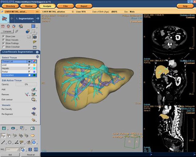
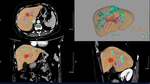
The CT Liver Analysis application provides a comprehensive analysis of the liver, allowing efficient patient management and treatment planning. Taking advantage of the power of the IntelliSpace Portal platform, the liver analysis application is an outstanding tool for extracting information from a liver CT, communicating this information to physicians and surgeons, planning the strategy for patient management, and for guiding surgery.
IntelliSpace Portal CT Liver Analysis application
How it works
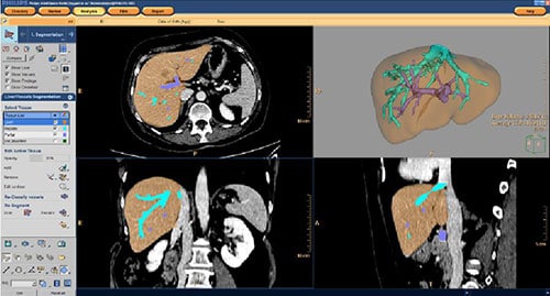
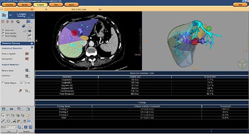
CT Liver Analysis provides Couinaud Lobe segmentation and interactive tools for planning liver resection and ablation. This robust application automatically identifies and extracts the liver using Enhanced Zero-click Performance. It then semi-automatically segments the liver for a comprehensive analysis and quantification of clinical informaiton. Once the segmentation is done, the resulting volume can be used for simple preplanning of the RF ablation of individual lesions or of partial liver resection.
For more information
Request a demonstration.
Download the Liver Analysis white paper.